Sonographer in the Making

Liver Flashcards

Informational, double sided. The most important facts on the card, an anatomy illustration and an ultrasound image on each, and a QR to the practice questions. No questions printed on the card.

Draft reformat of the original Liver cards. Content is Jenny's own; card size, front and back split, and QR targets are still to be set. Two cards shown; the full set follows the same pattern.
Card 1  ·  Liver: Anatomy
Front
SITM
LIVER
Anatomy
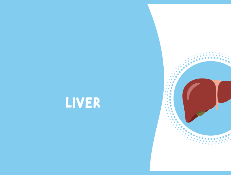
Liver illustration
Liver, anterior view

Key terms

  • Hepatocyte: liver cell performing all liver functions.
  • Falciform ligament: connects liver to diaphragm and abdominal wall; contains the ligamentum teres.
  • Ligamentum teres: remnant of the umbilical vein; bright echogenic focus; separates the medial and lateral segments of the left lobe.
  • Ligamentum venosum: separates the left lobe from the caudate lobe.
  • Main lobar fissure: boundary between right and left lobes; runs portal vein to gallbladder neck; landmark to the gallbladder.
  • Bare area: portion of liver with no peritoneal covering; against the diaphragm.

Position & lobes

  • Largest organ in the abdominal cavity; right hypochondrium, epigastrium, and left hypochondrium to the mammillary line.
  • Inferior to the diaphragm; right lobe covered by the ribs.
  • Lobes: right, left, and caudate (posterior lobe, independent vascular supply).
  • Variant: Riedel's lobe, a tongue like projection of the right lobe.
Practice QsQR
Back
SITM
LIVER
Vasculature
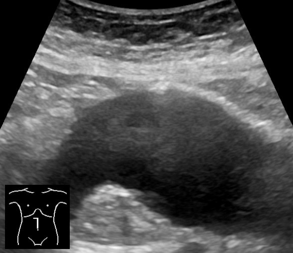
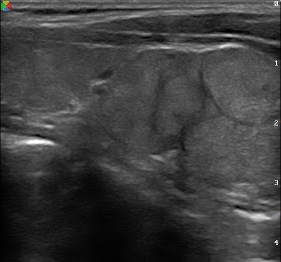
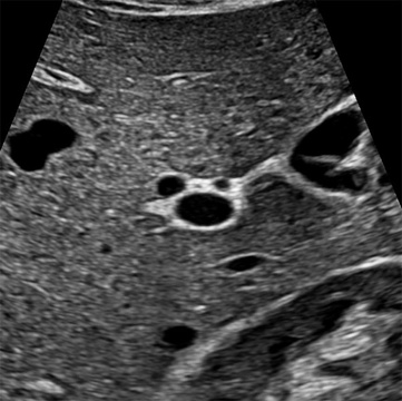
Transverse portal triad ultrasound
Portal triad, transverse: "Mickey Mouse sign"

Portal triad & blood supply

  • Portal triad: portal vein + hepatic artery + bile duct.
  • Mickey Mouse sign (transverse): portal vein is the face; hepatic artery and common bile duct are the two ears.
  • Vascular supply: portal vein 70 to 80% of blood; hepatic artery 20 to 30%, oxygenated.
  • Right lobe receives blood from the intestine; left and caudate lobes from the stomach and spleen.
  • Three hepatic veins (right, middle, left) drain into the IVC; portal vein enters at the porta hepatis.

Flow direction

  • Hepatopetal: flow toward the liver (normal portal flow).
  • Hepatofugal: flow away from the liver (normal hepatic vein flow; abnormal if portal).
Tricks: "petal" pulls toward  •  "fugitive" flees away

Embryology

  • Develops from the foregut endoderm; ligamentum teres is a remnant of the fetal umbilical vein.
Practice QsQR
Card 2  ·  Liver: Sonographic & Clinical
Front
SITM
LIVER
Sonographic
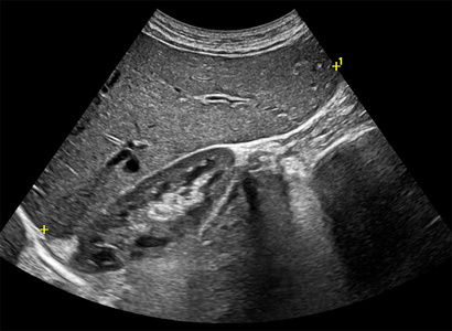
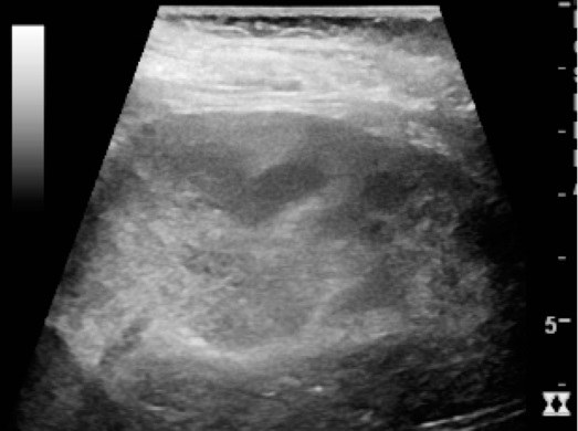
Right lobe echo texture ultrasound
Right lobe echo texture & measurement

Normal appearance

  • Homogeneous texture with fine, low level echoes.
  • Echogenicity: minimally hyperechoic to isoechoic vs renal cortex; hypoechoic vs spleen; pancreas equal to or slightly more echogenic than liver.
  • Portal veins have brighter (more echogenic) borders than hepatic veins (thicker collagen sheath). Ligaments and fissures appear echogenic to hyperechoic.

Measurements

  • TRV 21 to 22.5 cm; height 13 to 17.5 cm; AP depth 10 to 12.5 cm; SAG about 15.5 cm; portal vein 1.0 to 1.2 cm.
  • Longitudinal length over 20 cm indicates hepatomegaly.

Doppler

  • Hepatic veins show a normal triphasic waveform, reflecting right atrial hemodynamics.
  • Portal flow hepatopetal (toward liver); hepatic vein flow hepatofugal (away).
Practice QsQR
Back
SITM
LIVER
Function & Technique

Function

  • Metabolism: converts glucose to and from glycogen, processes fats and proteins.
  • Digestion: secretes bile, excretes bilirubin. Storage: iron and vitamins. Detoxification: breaks down drugs, ammonia, and toxins.

Lab values & clinical

  • AST / ALT high suggests hepatocellular damage. Alk Phos / direct bilirubin high suggests obstruction.
  • Ordered for abnormal LFTs, RUQ pain, jaundice, suspected hepatomegaly.
  • Before scanning, correlate liver echotexture vs kidney, spleen, and pancreas, and vascular patency.

Scanning & protocol

  • NPO 6 to 8 hrs; 2.5 to 5 MHz curvilinear or sector probe; supine or RAO, deep inspiration.
  • Survey 4 planes: sagittal, transverse, coronal, subcostal oblique. Measure portal vein at end inspiration.
  • Adequacy: about 15 cm (up to 15 to 20); homogeneous; liver brighter than kidney, less bright than pancreas and spleen; vessels, ligaments, and fissures visible; smooth surface; balanced gain (TGC).
Practice QsQR