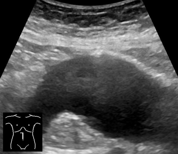
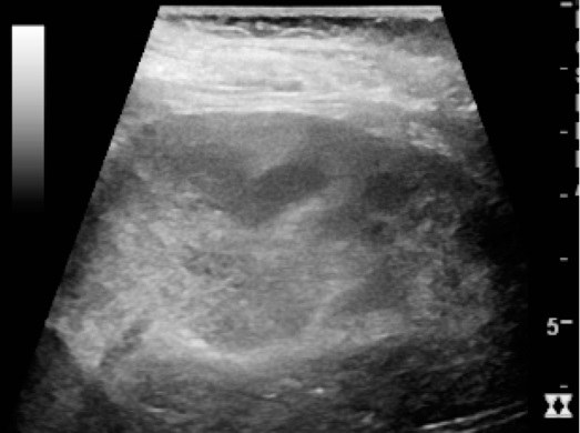
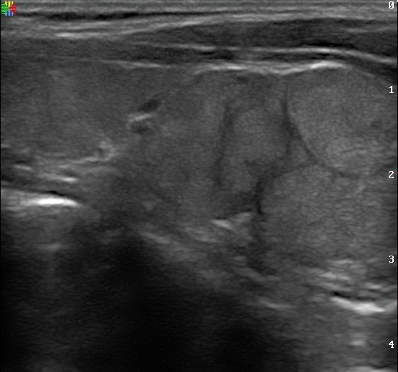
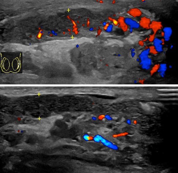
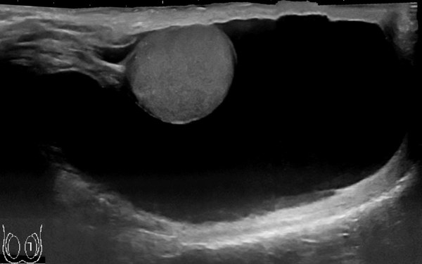
Sonographer in the Making

Abdomen & Small Parts Flashcards

Informational, double sided. The most important facts on the card, an anatomy illustration and an ultrasound image on each, and a QR to the digital practice questions. No questions printed on the card. Each topic opens with its normal card or cards, anatomy, function, labs, sonographic appearance, and scanning, then moves into the pathology.

33 double sided cards across 11 topics, in the order of your list: vascular system, liver, gallbladder and biliary, spleen, pancreas, gastrointestinal tract, peritoneal cavity and abdominal wall, urinary system, retroperitoneum, thyroid and parathyroid, scrotum. Still to come: breast and musculoskeletal if there is room, and the four topics you said could stay digital only, contrast agents, ultrasound guided intervention, emergent procedures, and the transplant patient. Content is grounded in the reference texts. Anatomy and ultrasound images are shown as labeled slots until the real figures are placed. Card size and QR targets still to be set.

Card 1  ·  Vascular System: Aorta & IVC
Front
SITM
VASCULAR
Anatomy
IllustrationAbdominal aorta with celiac trunk, SMA, renal arteries, IMA, and iliac bifurcation
Abdominal aorta and branch order

Course

  • Retroperitoneal structure lying anterior and slightly left of the spine
  • Continuation of the thoracic aorta, extending from the diaphragm to the common iliac artery bifurcation
  • Bifurcates at roughly the level of the umbilicus (near the fourth lumbar vertebra) into the right and left common iliac arteries
  • Tapers in diameter as it travels distally

Branches in order

  • Celiac trunk (first main visceral branch; gives rise to the splenic, common hepatic, and left gastric arteries)
  • Superior mesenteric artery (SMA), just inferior to the celiac trunk
  • Right and left renal arteries (right renal artery origin usually slightly superior to the left)
  • Inferior mesenteric artery (IMA)

Wall layers

  • Tunica intima (innermost, closest to flowing blood)
  • Tunica media (middle, muscular layer)
  • Tunica adventitia (outermost)
Practice QsQR
Back
SITM
VASCULAR
IVC & Scanning
UltrasoundTransverse image with pulsatile aorta left of spine and IVC to the right
Aorta and IVC in transverse

Inferior vena cava

  • Situated anterior to the spine and to the right of the aorta
  • Formed by the union of the common iliac veins posterior to the right common iliac artery
  • Major tributaries include the common iliac, renal, and hepatic veins (hepatic veins enter just below the diaphragm)
  • Left renal vein courses anterior to the aorta, posterior to the SMA, and enters the lateral wall of the IVC
  • Drains blood from the lower extremities and splanchnic territories into the right atrium
  • Travels a horizontal course with its proximal portion curving slightly anterior as it approaches the diaphragm

Aorta vs IVC

  • Aorta is pulsatile; IVC shows respirophasic variation and greater than 50 percent collapse with a sniff
  • Aorta lies to the left of midline; IVC lies to the right
  • IVC is thin walled and compressible; aorta is thick walled and non compressible

Scanning technique

  • Image the aorta in transverse and sagittal planes from the diaphragm to the bifurcation
  • Measure the anteroposterior diameter outer wall to outer wall, perpendicular to the vessel
  • Obtain measurements at the proximal, mid, and distal aorta and the proximal common iliac vessels
  • Normal adult male abdominal aorta is usually less than 3 cm in diameter
Practice QsQR
Card 2  ·  Vascular System: Portal Venous System
Front
SITM
VASCULAR
Portal System
IllustrationPortal vein formed behind the pancreas by the splenic and superior mesenteric veins, with its right and left branches in the liver
Portal venous system and its tributaries

Portal vein

  • Formed posterior to the pancreas by the union of the splenic vein and the superior mesenteric vein at about the level of L2
  • Trunk is roughly 5 to 7 cm long; it runs to the porta hepatis and divides into right and left portal veins
  • Carries blood from the intestinal tract to the liver, draining the gut from the lower esophagus to the upper anal canal, plus the pancreas, gallbladder, bile ducts, and spleen
  • Upper limit of normal for the main portal vein is about 13 mm anteroposterior; it varies with respiration

Dual blood supply

  • The liver is fed by both the portal vein and the hepatic artery
  • Portal blood is incompletely oxygenated, under 80 percent, yet supplies up to half the oxygen the hepatocytes need because its volume of flow is so large
  • The portal triad sits in a connective tissue sheath, which is what gives portal veins their bright echogenic walls
Practice QsQR
Back
SITM
VASCULAR
Tributaries & Findings
UltrasoundTransverse upper abdomen with the splenic vein crossing to meet the superior mesenteric vein at the portal splenic confluence
Portal splenic confluence in transverse

Splenic vein

  • Crosses the upper abdomen from the splenic hilum and is best seen in the transverse plane
  • Joins the superior mesenteric vein to form the main portal vein slightly to the right of midline

Superior mesenteric vein

  • Begins at the ileocolic junction and ascends to the right of the superior mesenteric artery
  • Passes anterior to the third part of the duodenum and posterior to the neck of the pancreas
  • Larger in caliber than the superior mesenteric artery, which arises directly off the anterior aortic wall
  • Forms the posterior border of the pancreatic neck and the anterior border of the uncinate process

Inferior mesenteric vein

  • Drains the left colon and ascends retroperitoneally along the left psoas to join the splenic vein behind the pancreas
  • Rarely identified sonographically: it is small, covered by small bowel, and has no posterior landmark to key on
Trick: Portal veins have bright walls, hepatic veins do not. Any large radicle near the porta hepatis is portal, not hepatic.
Practice QsQR
Card 3  ·  Vascular System: Abdominal Doppler
Front
SITM
VASCULAR
Flow Analysis
IllustrationSpectral Doppler display with time on the horizontal axis, velocity on the vertical axis, and a clear window under systole
Reading the spectral display

Must Know Terms

  • Resistive index
  • Spectral broadening
  • Plug flow
  • Sample volume
  • Hepatopetal
  • Hepatofugal

What Doppler answers

  • Whether flow is present, which direction it travels, and whether it is disturbed
  • Also used for tissue characterization and waveform analysis
  • Absence of flow separates the common bile duct from the hepatic artery beside it

Resistive vs nonresistive

  • Nonresistive vessels hold a high diastolic component and feed organs needing constant perfusion: internal carotid, hepatic artery, renal artery
  • Resistive vessels have little or even reversed diastolic flow and feed organs that do not need constant supply: external carotid, iliac, brachial
  • The resistive index compares peak systole against minimum diastole to quantify impedance

Reading the display

  • Horizontal axis is time; vertical axis is Doppler shift, so velocity. Flow toward the transducer sits above baseline, flow away sits below
  • Gray scale brightness shows how many cells are moving at a given velocity
  • Plug flow: most cells moving at the same velocity across the lumen, typical of large arteries, giving a clear window under systole
Practice QsQR
Back
SITM
VASCULAR
Technique & Patterns
UltrasoundTriphasic hepatic vein waveform with systolic, diastolic, and atrial components crossing the baseline
Triphasic hepatic vein waveform

Technique

  • Patient fasting, respiration suspended, done as part of the routine real time exam
  • Size the sample volume to the vessel and no wider. A gate wider than the lumen picks up neighbouring structures and produces noise and ghost echoes
  • Keep the beam parallel to flow. Accuracy holds to about 60 degrees; signal falls to zero at perpendicular, so roll the patient into obliquities rather than accept that angle

Arterial patterns

  • Aorta: proximal shows a high systolic peak with low diastole; the distal aorta turns triphasic as it nears the iliacs
  • Celiac axis: spectral broadening in diastole, unchanged after meals. The splenic artery is its most turbulent branch, from tortuosity
  • SMA: highly resistive fasting, with little or no diastolic flow, turning low resistance after a meal
  • Renal artery: nonresistive, with diastolic flow usually 30 to 50 percent of peak systole

Venous patterns

  • IVC and hepatic veins cross above and below baseline, reflecting right atrial reflux and respiration. Hepatic vein flow is triphasic
  • Portal flow is continuous, low velocity, and hepatopetal
  • Lost portal landmarks, a dilated splenic vein and SMV, and porta hepatis collaterals are indirect signs of portal vein thrombosis
Practice QsQR
Card 4  ·  Vascular System: Aortic Pathology
Front
SITM
VASCULAR
Aneurysm
UltrasoundSagittal abdominal aorta with focal fusiform dilatation and mural thrombus along the anterior wall
Abdominal aortic aneurysm with mural thrombus

Must Know Terms

  • Aneurysm
  • Fusiform
  • Saccular
  • Mural thrombus
  • Infrarenal
  • Outer to outer

Definition

  • An aneurysm is a permanent localized dilation of an artery, generally 1.5 times the normal vessel diameter or more
  • Focal dilatation of the abdominal aorta
  • Sonographically diagnosed when the abdominal aorta measures greater than 3 cm in diameter
  • Approximately 85 percent are infrarenal in location
  • Most occur at the level of the umbilicus, at or near the iliac bifurcation
  • Risk factors include atherosclerosis, hypertension, smoking, connective tissue disorders such as Marfan syndrome, and family history

Morphology

  • Fusiform: symmetric, circumferential (concentric) dilatation involving the full wall; most common shape and often contains mural thrombus
  • Saccular: focal outpouching involving only part of the wall; much less common and tends not to have thrombus deposition
  • Mural thrombus usually lies along the anterior or anterolateral wall of the lumen

Measurement

  • Measure the anteroposterior diameter on a longitudinal view, outer wall to outer wall
  • Measure the aorta at three levels (proximal, mid, distal) and at any suspected wall bulges
  • Include mural thrombus in the outer to outer measurement
Trick: If the lumen looks smaller than the outer wall, you are likely seeing mural thrombus; always place calipers outer wall to outer wall, not lumen to lumen.
Practice QsQR
Back
SITM
VASCULAR
Dissection & IVC
UltrasoundEnlarged aorta with a linear intimal flap dividing true and false lumens
Aortic dissection with intimal flap

Aortic dissection

  • Acute aortic syndrome characterized by an enlarged aorta with an intimal flap and demonstration of flow in a true and a false lumen
  • An intimal flap or membrane may be seen within the aortic lumen, with two patent channels or a thrombosed false lumen
  • May coexist with an underlying AAA
  • Clinical presentation includes sudden onset of severe chest pain with radiation to the arms, neck, or back; syncope may be present

IVC thrombus and tumor extension

  • Renal cell carcinoma commonly invades the renal vein; gross renal vein involvement occurs in about 21 to 35 percent of large tumors
  • A subset extend into the IVC and may reach the right atrium
  • Predisposing conditions include preexisting renal disease, hypercoagulable state, and IVC or ovarian vein thrombus with extension to the renal vein
  • Sonographic appearance: solid hypoechoic material within the IVC or renal vein lumen; assess for luminal filling defects and lack of compressibility
  • Evaluate with color and spectral Doppler to confirm absent or altered flow around the filling defect
Practice QsQR
Card 5  ·  Liver: Anatomy
Front
SITM
LIVER
Anatomy
Liver illustration
Liver, anterior view

Key terms

  • Hepatocyte: liver cell performing all liver functions.
  • Falciform ligament: connects liver to diaphragm and abdominal wall; contains the ligamentum teres.
  • Ligamentum teres: remnant of the umbilical vein; bright echogenic focus; separates the medial and lateral segments of the left lobe.
  • Ligamentum venosum: separates the left lobe from the caudate lobe.
  • Main lobar fissure: boundary between right and left lobes; runs portal vein to gallbladder neck; landmark to the gallbladder.
  • Bare area: portion of liver with no peritoneal covering; against the diaphragm.

Position & lobes

  • Largest organ in the abdominal cavity; right hypochondrium, epigastrium, and left hypochondrium to the mammillary line.
  • Inferior to the diaphragm; right lobe covered by the ribs.
  • Lobes: right, left, and caudate (posterior lobe, independent vascular supply).
  • Variant: Riedel's lobe, a tongue like projection of the right lobe.
Practice QsQR
Back
SITM
LIVER
Vasculature
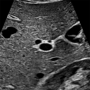
Transverse portal triad ultrasound
Portal triad, transverse: "Mickey Mouse sign"

Portal triad & blood supply

  • Portal triad: portal vein + hepatic artery + bile duct.
  • Mickey Mouse sign (transverse): portal vein is the face; hepatic artery and common bile duct are the two ears.
  • Vascular supply: portal vein 70 to 80% of blood; hepatic artery 20 to 30%, oxygenated.
  • Right lobe receives blood from the intestine; left and caudate lobes from the stomach and spleen.
  • Three hepatic veins (right, middle, left) drain into the IVC; portal vein enters at the porta hepatis.

Flow direction

  • Hepatopetal: flow toward the liver (normal portal flow).
  • Hepatofugal: flow away from the liver (normal hepatic vein flow; abnormal if portal).
Tricks: "petal" pulls toward  •  "fugitive" flees away

Embryology

  • Develops from the foregut endoderm; ligamentum teres is a remnant of the fetal umbilical vein.
Practice QsQR
Card 6  ·  Liver: Sonographic & Clinical
Front
SITM
LIVER
Sonographic
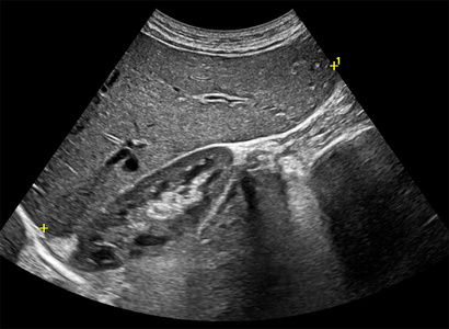
Right lobe echo texture ultrasound
Right lobe echo texture & measurement

Normal appearance

  • Homogeneous texture with fine, low level echoes.
  • Echogenicity: minimally hyperechoic to isoechoic vs renal cortex; hypoechoic vs spleen; pancreas equal to or slightly more echogenic than liver.
  • Portal veins have brighter (more echogenic) borders than hepatic veins (thicker collagen sheath). Ligaments and fissures appear echogenic to hyperechoic.

Measurements

  • TRV 21 to 22.5 cm; height 13 to 17.5 cm; AP depth 10 to 12.5 cm; SAG about 15.5 cm; portal vein 1.0 to 1.2 cm.
  • Longitudinal length over 20 cm indicates hepatomegaly.

Doppler

  • Hepatic veins show a normal triphasic waveform, reflecting right atrial hemodynamics.
  • Portal flow hepatopetal (toward liver); hepatic vein flow hepatofugal (away).
Practice QsQR
Back
SITM
LIVER
Function & Technique

Function

  • Metabolism: converts glucose to and from glycogen, processes fats and proteins.
  • Digestion: secretes bile, excretes bilirubin. Storage: iron and vitamins. Detoxification: breaks down drugs, ammonia, and toxins.

Lab values & clinical

  • AST / ALT high suggests hepatocellular damage. Alk Phos / direct bilirubin high suggests obstruction.
  • Ordered for abnormal LFTs, RUQ pain, jaundice, suspected hepatomegaly.
  • Before scanning, correlate liver echotexture vs kidney, spleen, and pancreas, and vascular patency.

Scanning & protocol

  • NPO 6 to 8 hrs; 2.5 to 5 MHz curvilinear or sector probe; supine or RAO, deep inspiration.
  • Survey 4 planes: sagittal, transverse, coronal, subcostal oblique. Measure portal vein at end inspiration.
  • Adequacy: about 15 cm (up to 15 to 20); homogeneous; liver brighter than kidney, less bright than pancreas and spleen; vessels, ligaments, and fissures visible; smooth surface; balanced gain (TGC).
Practice QsQR
Card 7  ·  Liver: Diffuse Disease  ·  Pathology
Front
SITM
LIVER
Diffuse Disease
Illustration Fatty (bright) liver vs. normal parenchyma
Diffuse fatty change

Fatty liver (steatosis)

  • Fatty deposits within the hepatocytes; the most common diffuse liver disease. Common causes: alcohol, obesity, diabetes.
  • Diffusely echogenic (bright) liver; increased sound beam attenuation, so the deep liver and diaphragm penetrate poorly and hepatic vessel walls are hard to see.
  • Focal fatty sparing, often near the gallbladder or porta hepatis, is a normal island in a bright liver and can mimic a mass.

Acute hepatitis

  • Inflammation of the liver. The parenchyma may look normal, or the portal vein borders become brighter than usual, the "starry sky" sign, with hepatomegaly.
Trick: bright liver that hides the diaphragm points to fat
Practice QsQR
Back
SITM
LIVER
Cirrhosis & Portal HTN
Ultrasound Cirrhotic liver: nodular surface, coarse echotexture, ascites
Cirrhosis with surface nodularity

Cirrhosis

  • End stage chronic liver disease; regenerating nodules replace normal parenchyma. Progression: steatosis to steatohepatitis to cirrhosis to portal hypertension.
  • Sonographic: shrunken, echogenic right lobe; enlarged caudate and left lobes; nodular surface irregularity (best seen against ascites or with a high frequency linear probe); coarse echotexture.
  • Clinical: hepatomegaly early, then jaundice, ascites, and splenomegaly.

Portal hypertension

  • Portal vein enlarges; portosystemic collaterals develop and the umbilical (paraumbilical) vein can recanalize; splenomegaly and ascites follow.
  • Flow: normal hepatopetal portal flow slows, becomes biphasic, then reverses to hepatofugal (away from the liver) in severe disease.
Trick: "petal" toward  •  "fugitive" away, so reversed portal flow (hepatofugal) is the red flag
Practice QsQR
Card 8  ·  Liver: Focal Lesions  ·  Pathology
Front
SITM
LIVER
Benign Lesions
Illustration Simple cyst and cavernous hemangioma
Common benign liver lesions

Simple hepatic cyst

  • Anechoic, round, smooth thin wall, with posterior acoustic enhancement (STAR criteria). Solitary or multiple.
  • Polycystic liver disease is associated with polycystic kidney disease.

Cavernous hemangioma

  • The most common benign tumor of the liver; a spongelike mass of blood filled spaces.
  • Typically well defined, homogeneous, and hyperechoic.

Other benign

  • Focal nodular hyperplasia and hepatic adenoma; adenoma is closely linked to oral contraceptive use.
Practice QsQR
Back
SITM
LIVER
Malignant & Infection
Ultrasound Hepatic metastases: multiple target lesions
Metastatic disease, target pattern

Hepatocellular carcinoma (HCC)

  • The most common primary liver malignancy; strongly linked to cirrhosis and chronic hepatitis.
  • Variable echogenicity; may invade the portal vein. Clinical: elevated AFP, weight loss.

Metastases

  • The liver is a common site for metastatic spread, and metastases are the most common malignant liver masses overall.
  • Multiple, variable echogenicity; may show a "target" or "bull's eye" pattern, an echogenic center with a hypoechoic halo.

Pyogenic abscess

  • A pus forming collection, often spread from appendicitis, diverticulitis, or cholecystitis.
  • Complex mass with internal echoes and debris; gas can cause dirty shadowing or ring down. Clinical: fever, hepatomegaly.
Practice QsQR
Card 9  ·  Gallbladder & Biliary: Normal
Front
SITM
GALLBLADDER
Anatomy & Function
IllustrationLabeled biliary tree: liver, right and left hepatic ducts joining as common hepatic duct, cystic duct with spiral valves of Heister, gallbladder fundus/body/neck, common bile duct entering duodenum
Gallbladder and biliary tree, labeled.

Regions of the Gallbladder

  • Fundus: rounded distal tip, projects beyond the inferior liver edge.
  • Body: mid portion, contacts the duodenum, transverse colon, and anterior abdominal wall.
  • Neck: narrow proximal end, continuous with the cystic duct.

Biliary Tree

  • Cystic duct: lined by the spiral valves of Heister, arising in the neck.
  • Common hepatic duct + cystic duct: unite to form the common bile duct (CBD).
  • CBD: joins the pancreatic duct at the ampulla of Vater to enter the duodenum.

Size & Blood Supply

  • Roughly 7 to 10 cm long, up to about 4 cm wide; holds approximately 30 to 50 mL of bile.
  • Blood supply: cystic artery (branch of the right hepatic artery); venous return via the cystic vein.

Anatomic Variants

  • Phrygian cap: fundus folds back on itself.
  • Junctional fold: kink at the neck.
  • Hartmann pouch: small outpouching near the neck where stones commonly lodge.

Function

  • Stores and concentrates bile produced by the liver.
  • Contracts in response to a fatty meal, releasing bile to the duodenum for fat emulsification.
Practice QsQR
Back
SITM
GALLBLADDER
Sonographic Appearance & Protocol
UltrasoundLong-axis grayscale image of a normal gallbladder: anechoic pear-shaped lumen with a thin echogenic wall, main lobar fissure pointing from the GB neck toward the right portal vein
Long axis, normal gallbladder along the main lobar fissure.

Normal Sonographic Appearance

  • Lumen: anechoic, pear-shaped, with posterior enhancement.
  • Wall thickness: less than 3 mm, measured on the anterior wall.
  • CBD diameter: up to about 6 mm in adults under 60; add roughly 1 mm per decade after 60, and up to about 10 mm may be normal post-cholecystectomy.
  • Landmark: the main lobar fissure connects the neck of the gallbladder to the right portal vein.

Scanning Protocol

  • Patient prep: NPO for at least 6 hours so the gallbladder is fully distended.
  • Transducer: broadband curvilinear, 2.5 to 5 MHz.
  • Position: begin supine; roll to left lateral decubitus or upright to shift stones and separate small stones from the wall.
  • Image and measure the gallbladder in long and transverse; measure wall on the transverse view, anterior wall, outer margin to outer margin, with the beam perpendicular.
Trick: Follow the main lobar fissure like an arrow; it points straight from the right portal vein to the gallbladder neck.
Practice QsQR
Card 10  ·  Gallbladder: Stones & Obstruction
Front
SITM
GALLBLADDER
Cholelithiasis & Sludge
UltrasoundGallbladder long axis with an echogenic stone in the dependent lumen casting a clean posterior acoustic shadow; second small panel showing gravity-dependent low-level echoes of sludge layering along the posterior wall
Gallstone with shadowing (left) and layering sludge (right).

Word Roots

cholelithiasis
bilestonecondition

Bile stone condition. Swap lith for cyst and you get cholecystitis.

Cholelithiasis

  • Classic triad: echogenic, mobile, posterior acoustic shadowing within the gallbladder lumen.
  • Stones shift to the most dependent portion when the patient rolls into left lateral decubitus or upright.
  • WES sign (Wall, Echo, Shadow): a contracted, stone-filled gallbladder shown as a bright wall, a curved echogenic arc, and dense posterior shadow.

Biliary Sludge

  • Low-level, nonshadowing echoes that layer in the dependent gallbladder.
  • Moves slowly with changes in patient position.
  • Tumefactive sludge: fills the lumen and can appear isoechoic to liver, mimicking a mass; still nonshadowing and mobile.
Trick: Stones shadow and shift; sludge layers and lags; polyps stay put and cast no shadow.
Practice QsQR
Back
SITM
GALLBLADDER
Choledocholithiasis & Obstruction
UltrasoundPorta hepatis long axis showing a dilated common bile duct anterior to the portal vein (parallel channel sign) with an echogenic shadowing stone within the distal CBD; small inset of dilated intrahepatic ducts running parallel to portal branches
Dilated CBD with an impacted stone; dilated duct paralleling the portal vein at the porta hepatis.

Choledocholithiasis

  • Stone within the common bile duct, often lodged at the distal CBD.
  • Sonographic appearance mirrors gallstones: echogenic focus within the duct with posterior shadowing.
  • CBD greater than about 6 mm is considered dilated in adults under 60; add roughly 1 mm per decade after 60, and up to about 10 mm may be normal post-cholecystectomy.

Biliary Obstruction

  • Bile ducts expand outward from the point of obstruction; extrahepatic dilation occurs before intrahepatic dilation.
  • Dilated CBD paralleling the portal vein at the porta hepatis (often called parallel channel sign).
  • Dilated intrahepatic ducts coursing parallel to portal vein branches within the liver (too many tubes / shotgun sign).
  • Note: terminology varies across texts; some sources apply parallel channel and shotgun sign to the extrahepatic finding, so favor the anatomic description.
  • Courvoisier gallbladder: painless jaundice with a distended, non-inflamed gallbladder from distal CBD obstruction, classically a pancreatic head carcinoma.
Trick: One tube next to the portal vein is normal; two parallel tubes is the CBD talking back.
Practice QsQR
Card 11  ·  Gallbladder: Wall & Inflammatory Pathology
Front
SITM
GALLBLADDER
Acute Cholecystitis
UltrasoundTransverse gallbladder with markedly thickened wall measured by calipers, gallstone impacted at the neck, and a rim of anechoic pericholecystic fluid tracking along the outer wall
Thickened gallbladder wall, impacted stone, and pericholecystic fluid.

Sonographic Findings

  • Wall thickening greater than 3 mm, measured on the anterior wall in transverse.
  • Distended lumen greater than 4 cm.
  • Gallstones, often with an impacted stone in Hartmann pouch or the cystic duct.
  • Pericholecystic fluid tracking along the gallbladder bed.

Sonographic Murphy Sign

  • Maximal tenderness reproduced when the transducer is pressed directly over the sonographically identified gallbladder.
  • Highly suggestive of acute cholecystitis when combined with wall thickening and stones.
  • May be diminished in advanced disease.
Trick: Think "3, 4, stone, sore": wall over 3 mm, lumen over 4 cm, stone impacted, positive Murphy under the probe.
Practice QsQR
Back
SITM
GALLBLADDER
Adenomyomatosis, Polyps & Porcelain GB
UltrasoundSplit image: focal fundal wall thickening with bright intramural foci throwing comet-tail artifact (adenomyomatosis) beside a non-mobile echogenic polyp attached to the anterior wall with no shadowing
Adenomyomatosis with comet-tail artifact and a wall-adherent polyp.

Adenomyomatosis

  • Benign hyperplasia of the gallbladder wall with Rokitansky-Aschoff sinuses.
  • Focal or diffuse wall thickening with small echogenic intramural foci.
  • Comet-tail artifact projecting from the wall into the lumen is characteristic.

Gallbladder Polyps

  • Echogenic soft tissue arising from the wall; most commonly cholesterol polyps.
  • Non-mobile with position change and produce no acoustic shadow.
  • May generate a comet-tail artifact, overlapping with adenomyomatosis.

Porcelain Gallbladder

  • Calcification of the gallbladder wall; typically clinically silent.
  • Wall appears as a bright echogenic curve with dense posterior shadowing, obscuring the lumen.
  • Associated with an increased risk of gallbladder carcinoma.
Practice QsQR
Card 12  ·  Gallbladder: Other Cholecystitis
Front
SITM
GALLBLADDER
Acalculous & Emphysematous
UltrasoundSplit panel: acalculous gallbladder with a thickened wall, layering sludge, and pericholecystic fluid but no stones in a critically ill patient; second panel showing bright echogenic foci within the gallbladder wall throwing dirty shadowing and ring-down artifact from intramural gas
Acalculous cholecystitis (left) and emphysematous cholecystitis with intramural gas (right).

Acalculous Cholecystitis

  • Cholecystitis without gallstones; seen in critically ill, trauma, burn, or post-op patients.
  • Sonographic findings mirror acute cholecystitis: wall thickening, sludge, pericholecystic fluid, positive sonographic Murphy, but no stones.
  • High morbidity; may progress rapidly to gangrene or perforation.

Emphysematous Cholecystitis

  • Gas within the gallbladder wall or lumen from gas-forming organisms; strongly associated with diabetes.
  • Bright echogenic foci with dirty shadowing and ring-down / reverberation artifact.
  • Gas rises to the non-dependent side and shifts with patient position, unlike stones.
Trick: No stones, sick patient, sick gallbladder equals acalculous; bright foci that rise instead of drop equals gas.
Practice QsQR
Back
SITM
GALLBLADDER
Gangrenous & Chronic
UltrasoundSplit panel: gangrenous gallbladder with a heterogeneous striated wall and intraluminal sloughed membranes; second panel showing a small contracted gallbladder wrapped around stones producing the wall-echo-shadow sign of chronic cholecystitis
Gangrenous cholecystitis with intraluminal membranes (left) and chronic cholecystitis with WES sign (right).

Gangrenous Cholecystitis

  • Advanced complication with wall necrosis; high risk of perforation.
  • Sonographic findings: heterogeneous or striated wall, intraluminal membranes from sloughed mucosa, focal wall defects.
  • Sonographic Murphy sign is often diminished or absent due to nerve damage.

Chronic Cholecystitis

  • Repeated inflammation from long-standing stones; wall becomes fibrotic and contracted.
  • Sonographic findings: small contracted gallbladder around stones, often producing the WES sign.
  • Wall thickening without acute inflammatory features (no pericholecystic fluid, negative Murphy).
Trick: Membranes and a quiet Murphy equals gangrene; small, stone-filled, silent equals chronic.
Practice QsQR
Card 13  ·  Spleen: Normal
Front
SITM
SPLEEN
Anatomy
IllustrationSpleen in LUQ, inferior to left hemidiaphragm, superolateral to left kidney, tail of pancreas at the splenic hilum
Spleen and neighbors, coronal view

Position & relations

  • Intraperitoneal organ of the left upper quadrant, just inferior to the diaphragm.
  • Left kidney lies inferior to the spleen.
  • Tail of the pancreas lies posterior to the stomach and lesser sac as it approaches the splenic hilum and splenic vessels.
  • A mass in the LUQ may displace the spleen inferiorly.

Function & parenchyma

  • Defense against disease (immune organ).
  • Hematopoiesis and erythropoiesis (active in the fetus).
  • Destruction and removal of flawed red blood cells and platelets.
  • Culling (destruction of aged or abnormal RBCs) and pitting (removal of intracellular inclusions from RBCs) occur in the red pulp.
  • Parenchyma is red pulp (venous sinuses, RBC turnover) plus white pulp (Malpighian corpuscles, the lymphoid follicles responsible for immune function).
  • Blood storage.

Variants: accessory spleen, polysplenia, asplenia

  • Accessory spleen (splenule): small round island of splenic tissue, typically located near the splenic hilum or near the tail of the pancreas.
  • Splenule appears isoechoic to the spleen on ultrasound.
  • Polysplenia: failure of fusion of splenic masses on the dorsal mesogastrium, producing multiple small spleens and often associated with cardiac or situs anomalies.
  • Asplenia: congenital absence of the spleen.
Trick: a splenule at the hilum can mimic a mass. Match its echotexture to the spleen to sort it out.
Practice QsQR
Back
SITM
SPLEEN
Sonographic & Technique
UltrasoundLongitudinal spleen with length measurement, upper margin near diaphragm to inferior margin
Longitudinal spleen with length measurement

Normal appearance

  • Homogeneous parenchyma; commonly isoechoic to slightly hyperechoic relative to the liver (references vary), and more echogenic than the left renal cortex.
  • Tubular structures visible at the splenic hilum.

Measurement

  • Measured on a longitudinal image from the upper margin (near the diaphragm) to the inferior margin.
  • Adult upper limit about 12 to 13 cm long axis; over 13 cm is splenomegaly; thickness over 6 cm also abnormal.
  • Compare parenchymal echogenicity to the liver on the same study.

Scanning technique

  • Coronal LUQ approach through intercostal spaces.
  • Right lateral decubitus opens the intercostal windows and brings the spleen into the coronal imaging plane; avoid steep decubitus, which can cause the spleen to fall away from the abdominal wall.
  • Deep inspiration brings the spleen into view.
  • Long axis of the spleen with color Doppler at the hilum; measure length. Transverse at the hilum; measure width.
Practice QsQR
Card 14  ·  Spleen: Pathology
Front
SITM
SPLEEN
Splenomegaly & Cyst
UltrasoundEnlarged spleen on longitudinal view with length caliper exceeding upper limit of normal
Splenomegaly, long axis

Mastery Check

  • Splenomegaly threshold
  • Congestive causes
  • Simple splenic cyst
  • Varices at the hilum

Fill in a circle when a topic feels solid.

Splenomegaly

  • Enlargement of the spleen beyond the upper limit of normal length on the longitudinal image.
  • Survey the parenchyma to confirm it remains uniform and homogeneous, aside from the tubular structures at the hilum.
  • When portal hypertension is suspected as the cause, closely evaluate the splenic hilum for abdominal varices.

Causes of congestive splenomegaly

  • Heart failure.
  • Portal hypertension, portal or splenic vein thrombosis.
  • Leukemia, lymphoma, mononucleosis.
  • Generalized infections, hemolytic anemias, glycogen storage disease.

Simple splenic cyst

  • Cystic masses are uncommon in the spleen; classified as congenital or acquired.
  • Sonographic findings: round, smooth walled, anechoic, with posterior enhancement.
Practice QsQR
Back
SITM
SPLEEN
Infarct & Trauma
UltrasoundPeripheral wedge shaped splenic infarct with base along the subcapsular surface
Splenic infarct, peripheral wedge

Splenic infarct

  • May show a localized hypoechoic area, depending on time of onset.
  • Fresh hemorrhage has a hypoechoic appearance.
  • Healed infarctions appear as echogenic, peripheral, wedge shaped lesions with their base toward the subcapsular surface.

Splenic trauma & rupture

  • After blunt injury a subcapsular hematoma may develop with subsequent rupture.
  • Assess for free fluid surrounding the splenic capsule in blunt abdominal trauma.
  • Small hypoechoic separation medial to the splenic capsule represents a subcapsular hematoma; inhomogeneity of the splenic texture may represent intraparenchymal hematoma.
  • Blood exhibits various echo patterns depending on the time since trauma.

Splenule vs mass

  • An accessory spleen is isoechoic to the spleen and typically sits at the hilum or near the pancreatic tail.
  • Match echotexture to the spleen to distinguish a splenule from a true splenic or peripancreatic mass.
Trick: splenosis (ectopic splenic implants) can follow splenic rupture and mimic peritoneal masses.
Practice QsQR
Card 15  ·  Pancreas: Normal
Front
SITM
PANCREAS
Anatomy & Landmarks
IllustrationTransverse pancreas with head, uncinate, neck, body, and tail draped over SMA, SMV, splenic vein, aorta, and IVC
Pancreas and its retroperitoneal vascular landmarks

Word Roots

pancreas
allflesh

Greek: "all flesh", named for its uniform fleshy texture.

Four Regions

  • Head: largest portion, seated in the C-loop of the duodenum to the right of the SMV
  • Uncinate process: posteromedial extension of the head that hooks behind the SMV
  • Neck: short segment anterior to the SMV and portal confluence
  • Body and tail: extend leftward toward the splenic hilum

Vascular Landmarks

  • Posterior: aorta and IVC are the posterior landmarks of the gland
  • Splenic vein: hugs the posterior border of the body and tail; primary sonographic landmark
  • SMA and SMV: lie posterior to the neck and body; SMA sits to the left of the SMV
  • CBD: courses through the posterolateral head
Practice QsQR
Back
SITM
PANCREAS
Duct, Echotexture & Function
UltrasoundTransverse epigastric image with splenic vein posterior to body and thin anechoic pancreatic duct centrally
Normal pancreatic duct with splenic vein landmark

Pancreatic Duct (Duct of Wirsung)

  • Normal duct measures less than 2 mm
  • Seen centrally within the body as two parallel echogenic walls with an anechoic lumen

Echogenicity

  • Normally isoechoic to hyperechoic relative to the liver
  • The pancreas is typically more echogenic than the adjacent liver parenchyma
  • Gland atrophies and becomes more echogenic with advancing age from fatty replacement

Function

  • Exocrine: acini cells secrete digestive enzymes; amylase digests carbohydrates and lipase digests fats
  • Endocrine: islets of Langerhans secrete insulin and glucagon into the blood

Scanning Approach

  • Transverse epigastric plane is the primary window
  • Head with IVC and SMV; body and tail with SMV and SMA
  • Water in the stomach can be used as an acoustic window when bowel gas obscures the gland
Practice QsQR
Card 16  ·  Pancreas: Inflammatory Pathology
Front
SITM
PANCREAS
Acute Pancreatitis
UltrasoundDiffusely enlarged hypoechoic pancreas with adjacent peripancreatic fluid
Diffuse acute pancreatitis with peripancreatic fluid

Sonographic Findings

  • Gland may appear normal early in the disease
  • Diffusely enlarged, hypoechoic pancreas from edema
  • Focal hypoechoic area within the gland in focal disease
  • Borders remain distinct but become irregular
  • Peripancreatic fluid collections; may progress to hemorrhage

Clinical & Labs

  • Elevated amylase within 24 hours
  • Elevated lipase within 72 hours; lipase is the more specific enzyme
  • Leukocytosis and elevated ALT may be present
  • Common causes include alcohol use, gallstones, and hypercalcemia
Practice QsQR
Back
SITM
PANCREAS
Pseudocyst & Chronic Pancreatitis
UltrasoundAnechoic pseudocyst arising from the pancreas with posterior enhancement
Pancreatic pseudocyst following acute pancreatitis

Pancreatic Pseudocyst

  • Complication of acute or chronic pancreatitis
  • Pancreatic juice escapes and collects in the anterior pararenal space
  • Typically anechoic with well-defined walls and posterior acoustic enhancement
  • May contain internal debris or septations after hemorrhage or infection
  • Extrapancreatic collections may resolve spontaneously within 4 weeks of onset

Pancreatic Abscess / Phlegmon

  • Complication of acute pancreatitis in which pancreatic enzymes accumulate in the surrounding space and become infected
  • Appears as a complex collection with internal debris, septations, and possible gas

Chronic Pancreatitis

  • Recurring destruction of pancreatic tissue leading to atrophy and fibrosis with scarring
  • Parenchymal calcifications within the gland cast posterior shadowing
  • Dilated pancreatic duct greater than 2 mm
  • Gland becomes small and heterogeneous
Practice QsQR
Card 17  ·  Pancreas: Neoplastic Pathology
Front
SITM
PANCREAS
Adenocarcinoma
UltrasoundHypoechoic poorly defined mass in the pancreatic head with irregular borders
Adenocarcinoma of the pancreatic head

Mnemonic

The three P's of a head mass: Painless jaundice, Palpable gallbladder, Pancreatic head. Painless is the word that separates it from stones.

Sonographic Findings

  • Most common appearance is a hypoechoic mass in the head of the pancreas
  • Poorly defined focal mass with an irregular border
  • Focal enlargement and displacement of normal pancreatic parenchyma
  • Secondary enlargement of the common duct from edema or tumor invasion of the head

Associated Signs

  • Double duct sign: coexisting dilation of the common bile duct and the pancreatic duct
  • Courvoisier gallbladder: painlessly enlarged palpable gallbladder caused by a pancreatic head mass obstructing the CBD
Practice QsQR
Back
SITM
PANCREAS
Adenocarcinoma: Clinical & Search Pattern
IllustrationPancreatic head mass with simultaneously dilated CBD and pancreatic duct, plus a distended gallbladder
Double duct sign with Courvoisier gallbladder

Clinical Presentation

  • Painless jaundice is the classic presentation of a head mass obstructing the CBD
  • Weight loss and anorexia are common
  • Ampullary adenocarcinomas carry a better prognosis than pancreatic adenocarcinoma

Search Pattern

  • Identify the focal hypoechoic mass and document its region: head, body, or tail
  • When the mass is in the head, look for CBD and pancreatic duct dilation
  • Evaluate the gallbladder for painless distention (Courvoisier)
  • Survey the liver for metastatic disease and assess regional lymph nodes
Trick: A hypoechoic head mass with the double duct sign and a distended, non-tender gallbladder should raise concern for pancreatic adenocarcinoma.
Practice QsQR
Card 18  ·  Gastrointestinal Tract: Normal
Front
SITM
GI TRACT
Anatomy
IllustrationStomach with fundus, body, and pylorus, the four duodenal segments, and the colon with its haustra
Regions of the gastrointestinal tract

Stomach and duodenum

  • Stomach in three parts: the fundus superiorly, the body forming the central axis, the pylorus distally
  • Duodenum in four segments: superior, descending, transverse, ascending

Small bowel and colon

  • Valvulae conniventes: large mucosal folds projecting into the small bowel lumen that slow the passage of food and increase absorption. Seen as linear echoes about 3 to 5 mm apart
  • Haustra: the sacculations that give the colon its segmented appearance
  • The vermiform appendix is a remnant of the apex of the cecum. It sits under McBurney point, the midpoint of a line from the right anterosuperior iliac spine to the umbilicus

Blood supply

  • The celiac, superior mesenteric, and inferior mesenteric arteries supply both small and large intestine
  • The celiac axis reaches the duodenum through its right gastric, gastroduodenal, and superior pancreaticoduodenal branches
  • Varices may arise from the gastroesophageal vessels
Practice QsQR
Back
SITM
GI TRACT
Wall & Technique
UltrasoundTransverse bowel loop showing the alternating echogenic and hypoechoic wall layers
The five layer bowel wall

Wall layers

  • The bowel wall has five layers. The odd numbered layers, first, third, and fifth, are echogenic; the even numbered layers, second and fourth, are hypoechoic
  • Average total thickness is about 3 mm when distended and 5 mm when undistended

Landmarks

  • The gastroesophageal junction is seen on the sagittal scan just left of midline as a target or bull eye, anterior to the aorta, posterior to the left lobe of the liver, and inferior to the hemidiaphragm
  • The duodenum outlines readily with water ingestion or a change in patient position

Technique and limits

  • Intraluminal air produces an echogenic shadow that stops the beam, which is why much of the tract is difficult to assess
  • The small bowel is usually not resolved beyond the valvulae conniventes
  • Digestion and absorption are the primary functions of the tract
  • Peristalsis is the discriminator: bowel changes shape under gentle transducer pressure while lymph nodes hold theirs
Practice QsQR
Card 19  ·  Gastrointestinal Tract: Upper Tract Pathology
Front
SITM
GI TRACT
Stomach
UltrasoundGastric wall thickening with a hypoechoic rim surrounding an echogenic lumen
Target appearance of gastric wall thickening

Gastric masses

  • Leiomyoma is the most common tumor of the stomach
  • Gastric carcinoma is the fifth leading cause of cancer and the third leading cause of cancer death
  • A polyp is a protruding, space occupying epithelial lesion within the stomach

Other gastric findings

  • Gastric bezoar: an intragastric mass made of accumulated ingested material
  • Duplication cyst criteria, all three required: lined with alimentary tract epithelium, a well developed muscular wall, and continuity with the stomach
Trick: A thickened hypoechoic wall around an echogenic lumen is the target or pseudokidney appearance. It flags bowel wall pathology without naming the cause.
Practice QsQR
Back
SITM
GI TRACT
Small Bowel
UltrasoundDilated fluid filled small bowel loops proximal to a point of obstruction
Dilated loops in small bowel obstruction

Obstruction

  • Small bowel obstruction shows dilation of the loops proximal to the site of obstruction
  • Fluid filled loops transmit sound, so an obstructed bowel is often easier to image than a normal one
  • Paralytic ileus dilates bowel without a mechanical obstructing point

Other lesions

  • Meckel diverticulum: a pouchlike herniation through the muscular wall of a tubular organ
  • Lymphomatous involvement of the intestinal wall can produce a pseudokidney or hydronephrotic pseudokidney appearance
  • Diverticulum: an outpouching of the wall, most often colonic
Practice QsQR
Card 20  ·  Gastrointestinal Tract: Appendix & Lower Tract
Front
SITM
GI TRACT
Appendicitis
UltrasoundNoncompressible blind ending tubular structure in the right lower quadrant with a thickened wall
Inflamed appendix on graded compression

Red Flag Features

  • Wall over 2 mm thick
  • Will not compress
  • Appendicolith with shadow
  • Asymmetric wall thickening
  • Surrounding free fluid

Acute appendicitis

  • Results from luminal obstruction and inflammation, leading to ischemia of the vermiform appendix
  • Presents with pain and rebound tenderness, usually localized over the right lower quadrant
  • Wall edema measures greater than 2 mm thick
  • Asymmetric wall thickening raises the possibility of perforation

Scanning it

  • High frequency linear transducer with graded compression over the point of maximum tenderness
  • A normal appendix compresses. An inflamed appendix does not
  • An appendicolith appears as an echogenic focus with posterior shadowing
  • Follow the blind ending tube to its tip; a normal tip does not exclude disease if the base was not seen
Trick: Compressibility is the whole test. If it flattens under the transducer, it is not an inflamed appendix.
Practice QsQR
Back
SITM
GI TRACT
Lower Tract
UltrasoundThickened terminal ileal wall with loss of the normal layered pattern
Bowel wall thickening in inflammatory disease

Inflammatory disease

  • Crohn disease is regional enteritis, a recurrent granulomatous inflammatory disease affecting the terminal ileum, the colon, or both, at any level
  • Inflamed segments show wall thickening with loss of the normal five layer pattern

Appendiceal and peritoneal

  • Mucocele of the appendix: gross enlargement of the appendix from accumulation of mucoid substance within the lumen
  • Pseudomyxoma peritonei: mucinous material distributed through the peritoneal cavity
Practice QsQR
Card 21  ·  Peritoneal Cavity & Abdominal Wall: Anatomy
Front
SITM
PERITONEUM
Anatomy
IllustrationSagittal section showing the greater sac, the lesser sac behind the stomach, and the subphrenic and subhepatic spaces
Peritoneal spaces in sagittal section

Two layers, two sacs

  • The parietal peritoneum lines the walls of the cavity; the visceral peritoneum covers the abdominal organs
  • The general peritoneal cavity is the greater sac
  • The lesser sac, or omental bursa, is the peritoneal recess posterior to the stomach
  • The cavity contains the greater and lesser omentum, the mesenteries, the ligaments, and the fluid spaces

Why the attachments matter

  • The peritoneal attachments to the walls and organs determine where abnormal fluid can collect and how it moves
  • Because of the coronary ligament attachments, a collection in the right posterior subphrenic space cannot extend between the bare area of the liver and the diaphragm
  • The bare area is delineated by the right superior and inferior coronary ligaments, which separate the posterior subphrenic space from the right superior subhepatic space, Morison pouch
  • Ligaments on the right of the liver form the subphrenic and subhepatic spaces
Practice QsQR
Back
SITM
PERITONEUM
Fluid & Collections
UltrasoundAnechoic free fluid outlining bowel loops in the dependent portion of the abdomen
Free fluid in the peritoneal cavity

Pelvic compartments

  • The retrovesical space is divided by the uterus into an anterior vesicouterine recess and a posterior rectouterine sac, the pouch of Douglas
  • The pouch of Douglas is the most dependent part of the peritoneal cavity in the supine patient, so free fluid gathers there

Ascites

  • Ascites is the accumulation of serous fluid in the peritoneal cavity
  • Simple ascites is anechoic and shifts with position; inflammatory or malignant ascites tends to carry internal echoes, septations, or matted bowel
  • The hepatorenal recess, Morison pouch, is a dependent space where small volumes collect first

Abscess

  • An abscess is a cavity formed by necrosis within solid tissue, or a circumscribed collection of purulent material
  • A gas containing abscess shows bright reflectors with dirty shadowing and reverberation
  • Named collections to look for: lesser sac, subphrenic, subcapsular, biloma
Practice QsQR
Card 22  ·  Peritoneal Cavity & Abdominal Wall: Pathology
Front
SITM
PERITONEUM
Mesentery & Omentum
UltrasoundHypoechoic band following the convexity of the anterior abdominal wall
Omental band in lymphoma

How masses behave

  • A mass in the mesentery or omentum may be cystic or solid; a mass within the peritoneum tends to show an infiltrative pattern
  • With an omental mass, at least one third are malignant, and secondary neoplasms outnumber primary
  • In the mesentery, a benign primary tumor is more common than a malignant one, secondary neoplasms still outnumber primary, and a cystic mass is more common than a solid one

Named lesions

  • Lymphoma presents as a uniformly thick, hypoechoic, band shaped structure following the convexity of the anterior and lateral abdominal wall, the omental band
  • Secondary tumors and lymphoma are the neoplasms that most commonly involve the peritoneum and mesentery
  • Urachal cyst: incomplete regression of the urachus during development
  • Urinoma: an encapsulated collection of urine from closed renal injury, surgery, or an obstructing lesion
Practice QsQR
Back
SITM
PERITONEUM
Abdominal Wall
UltrasoundHypoechoic collection within the rectus muscle sheath
Rectus sheath hematoma

Wall lesions

  • Lesions of the superficial abdominal wall include inflammatory lesions, hematomas, neoplasms, hernias, and postsurgical lesions
  • Rectus sheath hematoma: an acute or chronic collection of blood lying within the rectus muscle or between the muscle and its sheath
  • Lymphocele: a fluid collection appearing after surgery in the pelvis, retroperitoneum, or recess cavities
  • Neoplasms of the wall include lipomas, desmoid tumors, and metastases

Hernia

  • An abdominal hernia is the protrusion of a peritoneal lined sac through a defect in the weakened abdominal wall
  • Scan the defect at rest and during Valsalva, since a reducible hernia may only appear on strain
Trick: A hernia is a moving diagnosis. If the patient does not strain, a reducible sac stays hidden and the study reads normal.
Practice QsQR
Card 23  ·  Urinary System: Normal
Front
SITM
KIDNEY
Anatomy
IllustrationCoronal kidney showing cortex, medullary pyramids, columns of Bertin, renal sinus, capsule, and hilum
Renal anatomy

Location & Coverings

  • Retroperitoneal, paired organs
  • Three coverings from inner to outer: fibrous true capsule, perinephric (perirenal) fat, and Gerota's fascia
  • Hilum transmits renal artery, renal vein, and ureter

Parenchyma & Sinus

  • Parenchyma = renal cortex plus renal medulla
  • Medullary (renal) pyramids are triangular structures within the medulla
  • Columns of Bertin are cortical tissue extending between pyramids
  • Central renal sinus contains fat, calyces, pelvis, and vessels

Vasculature

  • Renal artery divides into about 5 segmental branches
  • Renal vein drains into the IVC

Function

  • Filter blood, produce urine, and maintain homeostasis
  • Urine drains calyces to renal pelvis to ureter to bladder
Practice QsQR
Back
SITM
KIDNEY
Sonographic Appearance
UltrasoundLong right kidney using liver as acoustic window with hypoechoic pyramids and echogenic central sinus
Normal long right kidney

Size

  • Normal adult kidney typically greater than 8 cm in longitudinal length
  • Average length 10 to 12 cm
  • Measure bipolar (pole to pole) long axis length

Echogenicity

  • Normal cortex is hypoechoic to or isoechoic with adjacent liver or spleen
  • Cortex should measure more than 1 cm in thickness
  • Medullary pyramids appear as triangular hypoechoic areas
  • Renal sinus is the brightest region (echogenic fat)

Scanning

  • Right kidney: use liver as an acoustic window
  • Left kidney: use spleen as an acoustic window
  • Slight decubitus rolls the liver anterior to the right kidney for better access
  • Document renal echogenicity compared with liver and spleen

Bladder

  • Anechoic when distended with homogeneously echogenic wall
  • Wall uniform in thickness, less than 3 mm when well distended
  • Color Doppler shows ureteral jets entering from the trigone at the posterolateral bladder base

Renal Function Labs

  • BUN and creatinine are the standard renal function labs
  • Both are elevated in renal failure
Practice QsQR
Card 24  ·  Urinary System: Cystic & Obstructive
Front
SITM
KIDNEY
Hydronephrosis
UltrasoundLong kidney with anechoic dilated central collecting system separating the calyces
Dilated collecting system

Definition

  • Dilation of the renal collecting system
  • Formal systems include SFU Grades I to IV and RAD I to V; also described descriptively as mild, moderate, severe

Sonographic Findings

  • Anechoic distension of the renal pelvis and calyces
  • Mild: distension of the renal pelvis
  • Moderate: pelvis and calyces dilate further with ballooning
  • Severe: marked dilation with cortical thinning

Simple Renal Cyst (Bosniak I)

  • Round or oval, anechoic with no internal echoes
  • Thin, well-defined walls with sharp interface to parenchyma
  • Posterior acoustic enhancement (through transmission)
  • No color flow within the mass
Practice QsQR
Back
SITM
KIDNEY
ADPKD
UltrasoundEnlarged kidney with numerous bilateral cortical cysts replacing much of the parenchyma
ADPKD

Autosomal Dominant Polycystic Kidney Disease

  • Previously known as adult polycystic kidney disease
  • Presents with hypertension, flank pain, and progressive renal failure

Sonographic Findings

  • Bilateral enlarged kidneys containing numerous cortical renal cysts
  • Cysts vary in size and may distort the normal renal contour
  • Associated hepatic cysts may be present

Distinguishing from Simple Cysts

  • Multiple bilateral cysts with kidney enlargement, not a solitary lesion
  • Progressive replacement of normal parenchyma
  • Screen the liver for associated cysts
Trick: Bilateral, numerous, and enlarged kidneys with liver cysts points to ADPKD rather than incidental simple cysts.
Practice QsQR
Card 25  ·  Urinary System: Masses & Stones
Front
SITM
KIDNEY
Urolithiasis
UltrasoundEchogenic focus within the renal sinus casting a clean posterior acoustic shadow
Renal calculus with shadowing

Clinical

  • Renal colic, hematuria, oliguria, urinary tract infection

Sonographic Findings

  • Echogenic focus that produces posterior acoustic shadowing
  • Twinkle sign seen posterior to the calculus on color Doppler
  • May cause upstream hydronephrosis if obstructing

Pitfall

  • Prominent renal sinus fat, mesenteric fat, and bowel gas can appear as an indistinct echogenic focus with questionable shadowing
  • Twinkle artifact helps confirm a true stone
Practice QsQR
Back
SITM
KIDNEY
Solid Renal Masses
UltrasoundSolid renal mass distorting the cortical contour with internal color Doppler flow
Solid renal mass

Renal Cell Carcinoma

  • Solid mass on the kidney, hypoechoic, isoechoic, or hyperechoic
  • Can have a complex cystic appearance
  • Consider RCC, oncocytoma, angiomyolipoma, transitional cell carcinoma, and secondary neoplasms when a solid mass is detected

Angiomyolipoma

  • Benign fat-containing tumor of the kidney
  • Classically well defined and markedly hyperechoic (fat)
  • A small hyperechoic RCC can mimic it

Mimic to Exclude

  • Hypertrophied column of Bertin is a normal variant that can mimic a mass
  • Confirm continuity with adjacent cortex and matching echogenicity
Trick: A well-defined hyperechoic renal mass suggests angiomyolipoma, but RCC can also appear hyperechoic, so correlate and follow up.
Practice QsQR
Card 26  ·  Retroperitoneum: Spaces & Adrenal Anatomy
Front
SITM
RETROPERITONEUM
Spaces
IllustrationAxial section showing the anterior pararenal, perirenal, and posterior pararenal spaces around the kidney
The three retroperitoneal compartments

Boundaries

  • The retroperitoneal space lies between the posterior parietal peritoneum and the posterior abdominal wall muscles, extending from the diaphragm to the pelvis
  • Laterally it reaches the extraperitoneal fat planes within the transversalis fascia; medially it encloses the great vessels

Three compartments

  • Perirenal space: the kidney, the adrenal, and perirenal fat
  • Anterior pararenal space: the duodenum, the pancreas, and the ascending and transverse colon
  • Posterior pararenal space: the iliopsoas muscle, the ureter, and branches of the IVC and aorta with their lymphatics

Node bearing areas

  • Two major lymph node bearing areas: the iliac and hypogastric nodes within the pelvis, and the para-aortic group in the upper retroperitoneum
  • The prevesical space runs from the pubis to the anterior margin of the bladder; the presacral space lies between the rectum and the fascia over the sacrum
Practice QsQR
Back
SITM
RETROPERITONEUM
Adrenal Anatomy
UltrasoundLongitudinal right upper quadrant view through the liver showing the adrenal region above the upper pole of the right kidney
Right adrenal region through the liver

Position and shape

  • The right adrenal sits more superior to the kidney; the left sits more medial to it
  • The right adrenal has a comma or triangular shape in the transaxial plane
  • A single vein drains each gland: the right into the IVC, the left into the left renal vein

How to find them

  • Right: scan longitudinally through the right lobe of the liver, perpendicular to the linear right crus of the diaphragm
  • Left: harder because of stomach gas. Place the patient right lateral decubitus, scan along the posterior axillary line using the spleen and left kidney as a window, in deep inspiration
  • Recognize retroperitoneal fat as separate from the liver, the crus, the gland, and the great vessel

Pitfalls that mimic the gland

  • Right crus of the diaphragm, the second portion of the duodenum, the gastroesophageal junction, medial lobulations of the spleen, splenic vasculature, the body and tail of the pancreas, and the fourth portion of the duodenum
Practice QsQR
Card 27  ·  Retroperitoneum: Adrenal Pathology
Front
SITM
RETROPERITONEUM
Cortical Syndromes
IllustrationAdrenal gland in cross section with the outer cortex and the inner medulla labeled
Adrenal cortex and medulla

What each part secretes

  • Cortex: mineralocorticoids, chiefly aldosterone, which regulate electrolyte and water balance; glucocorticoids, chiefly cortisol, which drive carbohydrate metabolism and damp inflammation; and small amounts of androgens and estrogens
  • Medulla: epinephrine and norepinephrine
  • The cortex is controlled by ACTH from the pituitary

The syndromes

  • Addison disease: adrenocortical insufficiency with cortical atrophy. Hypotension, weakness, fatigue, loss of appetite and weight, and a characteristic bronzing of the skin. Prognosis is good on steroid replacement
  • Conn syndrome: aldosteronism from excess aldosterone, present in about 0.5 percent of patients with sustained hypertension, usually from a cortical adenoma measuring 0.5 to 3 cm with contralateral adrenal atrophy. Muscle weakness, hypertension, abnormal electrocardiogram
  • Adrenogenital syndrome: excess sex hormones and adrenal androgens, from a tumor or from hyperplasia, producing virilization
Practice QsQR
Back
SITM
RETROPERITONEUM
Adrenal Masses
UltrasoundRounded solid mass superior to the upper pole of the kidney, separate from renal parenchyma
Suprarenal mass above the upper pole

Tumors

  • Benign nonfunctioning adenoma is the most common primary adrenal tumor
  • The adrenal glands are the fourth most common site in the body for metastasis, after the lung, the liver, and the bones
  • Pheochromocytoma arises from the pheochromocytes of the medulla and secretes epinephrine and norepinephrine in excessive quantities
  • Neuroblastoma is the most common adrenal malignancy of childhood and the most common tumor of infancy, representing about 30 percent of all neonatal tumors

Non neoplastic

  • Adrenal hemorrhage is most common in neonates after a traumatic delivery with stress, asphyxia, or septicemia. The gland may return to normal size with focal areas of calcification
  • Adrenal cysts appear as anechoic lesions with through transmission in the suprarenal location
Trick: A suprarenal mass is not automatically adrenal. Prove it is separate from the kidney, the crus, and the bowel before naming it.
Practice QsQR
Card 28  ·  Retroperitoneum: Nodes, Masses & Collections
Front
SITM
RETROPERITONEUM
Lymph Nodes
UltrasoundEcho poor rounded masses along the anterior and lateral margins of the aorta with the vessel displaced anteriorly
Para-aortic lymphadenopathy

Where and how to look

  • The nodes lie along the lateral and anterior margins of the aorta and the IVC, so scan supine or decubitus
  • Always examine in two planes; enlarged nodes should be reproducible in both projections
  • A left coronal view using the left kidney as a window reaches the para-aortic group

The patterns

  • Rounded, focal, echo poor lesions, 1 to 3 cm and larger
  • Confluent echo poor masses, which often displace the kidney laterally
  • A mantle of nodes in the paraspinal location
  • A floating aorta, displaced anteriorly by the enlarged nodes
  • The mesenteric sandwich sign, anterior and posterior nodal masses surrounding the mesenteric vessels

Node or bowel

  • Nodes hold their shape under gentle transducer pressure and transmit sound homogeneously
  • Bowel changes with peristalsis, shows dense central mucosal echoes, and often shadows from air within the wall
Trick: Use color flow before calling a lesion a node. A vessel in cross section looks exactly like one.
Practice QsQR
Back
SITM
RETROPERITONEUM
Masses & Collections
UltrasoundHypoechoic sheet of tissue enveloping the aorta and IVC anterior to the spine
Retroperitoneal fibrosis around the great vessels

Tumors

  • Lymphoma is the most common primary retroperitoneal tumor
  • Metastatic disease can occur anywhere in the retroperitoneum, arriving hematogenously, through the lymphatics, or by direct extension
  • Evaluate splenic size and the splenic hilum in any patient with lymphadenopathy

Fluid collections

  • Urinoma: a walled off collection of extravasated urine, developing spontaneously or after trauma, surgery, or subacute to chronic urinary obstruction
  • Retroperitoneal hemorrhage: seen with trauma, vasculitis, bleeding diathesis, a leaking aortic aneurysm, or a bleeding neoplasm

Fibrosis

  • Retroperitoneal fibrosis, Ormond disease, is an idiopathic condition characterized by thick sheets of fibrous tissue in the retroperitoneal cavity
  • It encases the great vessels and the ureters, so hydronephrosis is often the presenting finding
Practice QsQR
Card 29  ·  Thyroid & Parathyroid: Normal
Front
SITM
THYROID
Anatomy & Sonographic Appearance
IllustrationRight and left lobes joined by isthmus, trachea medial, CCA and IJV lateral, longus colli posterior
Thyroid gland in the anteroinferior neck

Location & Shape

  • Located in the anteroinferior neck, inferior to the thyroid cartilage
  • Right and left lobes connected across the midline by the isthmus
  • The lobes are normally equal in size, with a wide range of variability; a pyramidal lobe (superior extension of the isthmus) is a normal variant
  • Isthmus normally measures between 2 and 6 mm in the AP dimension

Sonographic Appearance

  • Fine, homogeneous echotexture
  • Slightly more echogenic than the surrounding musculature
  • Thin echogenic thyroid capsule

Transverse Landmarks

  • Trachea in the midline, posterior to the isthmus
  • Common carotid artery and internal jugular vein lateral to each lobe
  • Longus colli muscle posterior to the thyroid
  • Strap muscles (sternohyoid, sternothyroid, thyrohyoid, omohyoid) anterior; sternocleidomastoid anterolateral
Practice QsQR
Back
SITM
THYROID
Technique, Physiology & Parathyroid
IllustrationSupine patient with pillow under shoulders and neck hyperextended; four parathyroid glands posterior to thyroid
Scanning setup and parathyroid location

Patient & Transducer

  • Supine with a pillow or pad under both shoulders for moderate neck hyperextension
  • High-frequency linear array transducer, 7 to 15 MHz
  • Select the highest frequency that still penetrates
  • Scan each lobe in transverse and sagittal planes

Thyroid Physiology & Labs

  • Follicular cells produce thyroxine (T4) and triiodothyronine (T3); iodine required
  • Parafollicular (C) cells secrete calcitonin
  • TSH from the pituitary regulates thyroid hormone output
  • Labs typically show elevated TSH with low T3 and T4 in primary hypothyroidism, and the opposite pattern in hyperthyroidism

Parathyroid Glands

  • Endocrine glands located on the posterior surface of the thyroid
  • Most people have four parathyroid glands, though 3 to 5 glands is not uncommon
  • Normal parathyroid gland measures about 5 to 6 mm
  • Secrete parathyroid hormone (PTH), the principal regulator of calcium homeostasis
  • Normal glands are usually too small to identify on ultrasound
Trick: Thyroid is more echogenic than the adjacent strap muscles. If the gland looks darker than the straps, think diffuse thyroid disease.
Practice QsQR
Card 30  ·  Thyroid: Diffuse Disease
Front
SITM
THYROID
Goiter & Graves Disease
UltrasoundDiffusely enlarged thyroid with markedly increased color Doppler flow (thyroid inferno)
Graves disease with hypervascular gland

Confidence

ConfusedGot it

Shade one more block each time you review.

Goiter

  • Diffuse enlargement of the thyroid gland, with or without palpable nodules
  • Isthmus exceeding 1 cm in the AP plane suggests enlargement
  • Diffusely heterogeneous echotexture; may show focal scarring, ischemia, necrosis, or cyst formation
  • Multinodular goiter is a common cause of diffuse enlargement

Graves Disease

  • Most common cause of hyperthyroidism; autoimmune, more common in women
  • Diffusely enlarged, hyperplastic gland
  • Marked hypervascularity on color Doppler termed the "thyroid inferno"
  • Labs: high T3 and T4, low TSH
Practice QsQR
Back
SITM
THYROID
Hashimoto Thyroiditis
UltrasoundHeterogeneous, irregular thyroid with multiple ill-defined hypoechoic regions separated by echogenic bands
Chronic lymphocytic (Hashimoto) thyroiditis

Hashimoto Thyroiditis

  • Chronic lymphocytic thyroiditis; autoimmune, most common cause of hypothyroidism
  • More common in women
  • Mild enlargement of the gland initially, with later atrophy
  • Diffusely heterogeneous, hypoechoic echotexture
  • Multiple, ill-defined hypoechoic regions separated by echogenic fibrous bands

Clinical Correlation

  • Preexisting Hashimoto disease is a known risk factor for thyroid lymphoma
  • Labs typically show low T3 and T4 with elevated TSH
Trick: Both Graves and Hashimoto can look diffusely hypoechoic. Add color Doppler: Hashimoto vascularity is variable and typically less than the Graves inferno.
Practice QsQR
Card 31  ·  Thyroid: Nodules & Parathyroid
Front
SITM
THYROID
Nodules & Papillary Carcinoma
UltrasoundSolitary solid markedly hypoechoic thyroid nodule, taller-than-wide, with internal microcalcifications
Suspicious thyroid nodule

Red Flag Features

  • Taller than wide
  • Microcalcifications
  • Markedly hypoechoic
  • Irregular margins
  • Marked internal flow

Nodule Basics

  • Nodules may be cystic, solid, or complex
  • Simple anechoic cysts and colloid cysts (with echogenic focus and comet-tail) are typically benign
  • Follicular adenoma is the most common benign thyroid neoplasm; well-encapsulated solid mass
  • Any suspicious cystic or complex mass may require FNA biopsy

Suspicious (Malignant) Features

  • Solitary, solid mass that is markedly hypoechoic compared with the gland
  • Taller-than-wide shape
  • Internal microcalcifications (psammoma bodies)
  • Irregular margins
  • Marked internal vascularity

Papillary Carcinoma

  • Most common thyroid malignancy, approximately 70% of thyroid cancers
  • Classically hypoechoic solid mass with microcalcifications
  • May spread to cervical lymph nodes
Practice QsQR
Back
SITM
PARATHYROID
Parathyroid Adenoma
UltrasoundOval hypoechoic solid mass posterior to the lower pole of the thyroid
Parathyroid adenoma

Parathyroid Adenoma

  • Benign solid mass and the most common cause of primary hyperparathyroidism
  • Appears as a hypoechoic mass adjacent to or posterior to the thyroid gland
  • Usually solitary; most often near the lower pole
  • Normal parathyroid glands are typically not visualized

Primary Hyperparathyroidism

  • Elevated serum calcium
  • Elevated PTH
  • Most often caused by a parathyroid adenoma; less commonly parathyroid hyperplasia or, rarely, parathyroid carcinoma

Secondary Hyperparathyroidism

  • Associated with chronic renal failure and vitamin D deficiency
  • Elevated PTH with low calcium
  • May demonstrate enlargement of all four parathyroid glands
Trick: A hypoechoic mass hugging the posterior thyroid in a patient with high calcium and high PTH is a parathyroid adenoma until proven otherwise.
Practice QsQR
Card 32  ·  Scrotum & Testes: Normal
Front
SITM
SCROTUM
Anatomy
IllustrationTestis with mediastinum, tunica albuginea and vaginalis, epididymal head, body, and tail
Scrotal and testicular anatomy

Testis

  • Paired ovoid organs suspended within the scrotum
  • Covered by the tunica albuginea, a dense fibrous capsule
  • Tunica vaginalis is a double serous layer (parietal and visceral) covering the testis and epididymis; a potential space between the layers is where hydroceles form
  • Mediastinum testis appears as an echogenic band located along the posterolateral aspect of the testis

Epididymis and vas deferens

  • Three parts: head, body, and tail
  • Head lies superior to the upper pole of the testis; body and tail course posteriorly along the testis
  • Echogenicity is similar to or slightly less than the adjacent testis
  • The epididymal tail continues as the vas (ductus) deferens

Scrotal wall and spermatic cord

  • Dartos muscle divides the scrotum into two compartments; the cremaster muscle provides thermoregulation
  • Spermatic cord contents: vas deferens, testicular artery, cremasteric artery, artery to the vas, pampiniform plexus, lymphatics, and nerves

Vascular supply

  • Testicular artery branches into capsular artery, then centripetal artery, then recurrent rami
  • Cremasteric and deferential arteries accompany the testicular artery within the spermatic cord
Practice QsQR
Back
SITM
SCROTUM
Sonographic Technique
UltrasoundTransverse dual image of both testes with color Doppler for side by side comparison
Bilateral comparison view

Transducer

  • High frequency linear array transducer is preferred for scrotal imaging
  • Very high frequency probes (7 to 15 MHz) are typical for smaller superficial structures such as the scrotum

Patient and protocol

  • Patient is imaged supine with the scrotum supported
  • Each testis is evaluated in transverse and sagittal planes
  • Obtain a transverse image of the right and left testes together for side by side comparison in both gray scale and color Doppler
  • Include images of the epididymal head superior to the testis and follow the body and tail posteriorly

Doppler evaluation

  • Color and pulsed Doppler flow within each testicle and epididymis should be symmetric
  • Match color and spectral settings on the contralateral side before comparing
  • Perform the Valsalva maneuver when a varicocele is suspected
Practice QsQR
Card 33  ·  Scrotum & Testes: Pathology
Front
SITM
SCROTUM
Inflammatory & Torsion
UltrasoundColor Doppler of enlarged hypervascular epididymis versus opposite testis with absent flow
Hyperemia versus absent flow

Epididymitis and epididymo-orchitis

  • Most common cause of acute scrotal pain in adults
  • Inflamed epididymis appears enlarged, hypoechoic, or heterogeneous
  • Hyperemic flow within the epididymis and/or testis on color Doppler
  • Associated findings include a hypoechoic testis with orchitis, thickened scrotal wall, and reactive hydrocele

Testicular torsion

  • Most common cause of acute scrotal pain in adolescents
  • Early stages may show a normal sonographic appearance
  • After 4 to 6 hours the testis becomes swollen and hypoechoic
  • Color Doppler shows decreased or absent intratesticular flow compared with the contralateral testis
  • Paratesticular flow may be increased around the abnormal testis
Trick: Always set color Doppler on the asymptomatic side first, then apply identical settings to the painful side so a true flow difference is not hidden by scanner adjustments.
Practice QsQR
Back
SITM
SCROTUM
Fluid, Vascular & Mass
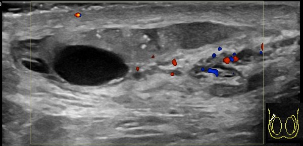
UltrasoundAnechoic fluid surrounding testis, serpiginous veins over 3 mm, hypoechoic intratesticular mass, echogenic foci
Extratesticular and intratesticular findings

One Root, Four Words

-celeswelling or hernia
  • hydro water, so hydrocele is clear fluid
  • hemato blood, so hematocele is blood
  • pyo pus, so pyocele is infection
  • varico dilated vein, so varicocele is veins

Hydrocele, hematocele, and pyocele

  • Hydrocele is a fluid filled collection located between the two layers of the tunica vaginalis, sonographically anechoic surrounding the testis
  • Hematocele = blood, pyocele = pus, in the same potential space; usually complex with septations and debris

Varicocele

  • Caused by incompetent valves within the pampiniform plexus
  • Veins measuring 3 mm or larger in diameter are considered varicose
  • Occurs most often on the left side
  • Distension increases with the Valsalva maneuver

Testicular carcinoma

  • Most testicular tumors are well defined hypoechoic intratesticular masses
  • Larger tumors may be poorly marginated or appear heterogeneous

Testicular microlithiasis

  • Multiple bright, nonshadowing echogenic foci scattered throughout the testis
Practice QsQR